Traditional microscopy is now a thing of the past. Artificial Intelligence (AI) and all its implications of machine learning are poised to revolutionize the way in which Pathology will be viewed and defined. With the conventional modes of diagnosis being replaced by technologically advanced digital tools and software opens up a plethora of opportunities toward medical advancement. With larger volumes of data, newer technological methods and endless resources that are available to pathologists at lower costs, AI in pathology is set to achieve many laurels that were previously considered impossible. Lab testing with artificial intelligence translates into intelligent algorithms that are capable of identifying general and specific patterns that can then offer logical and reliable predictions toward patient diseases.
Pathology in the ‘digital’ world further allows long distance association. This means that sending digital material from one end of the world to another comes bereft of constraints such as tampering. Through this association, the possibilities of collaboration are extended and lab growth is enhanced through increased access to specialists around the world. However, artificial intelligence in digital pathology cannot be looked at as a replacement of pathologists. Intelligent technologies, artificial intelligence for medical tests and AI-based diagnostics in healthcare are only a tool to increase the efficiency of human pathologists and cannot be looked at as an individual entity.
Pain-points in the current pathology scenario:
- Outsourcing of lab testing – The increased susceptibility of tampering due to the outsourcing culture established among pathology.
- Marginalization of pathologists in healthcare – Pathologists are increasingly disconnected from routine healthcare business practices and patients, causing isolated diagnosis.
- Reducing number of pathologists – The number of professionals going into the field of pathology is steadily declining.
- Complications in laboratory testing – The requirement of expertise and testing protocols create various complications in the lab testing procedures.
- Minimal clinical interaction – Pathologists are not involved in clinical processes and patient examination, which interferes with holistic diagnosis.
- Financial constraints – The financial constraints that come with lab testing hampers the quality of pathology.
- Resistance to new technologies – Most pathologists are more comfortable with conventional procedures and demonstrate a resistance to evolving technologies and machine learning.
The Traditional Pathology Process:
According to the National Cancer Institute, “A pathology report is a document that contains the diagnosis determined by examining cells and tissues under a microscope. The report may also contain information about the size, shape, and appearance of a specimen as it looks to the naked eye. This information is known as the gross description.
A pathologist is a doctor who does this examination and writes the pathology report. Pathology reports play an important role in cancer diagnosis and staging (describing the extent of cancer within the body, especially whether it has spread), which helps determine treatment options.”
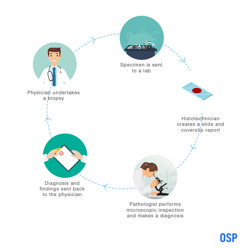
The conventional method of pathology begins when a physician or a clinician performs a biopsy on a patient and sends these samples to a pathological lab for testing. Let’s pursue this through an example. John Smith approaches a clinic with the complaint of a tumour in his left leg. The physician then prescribes for a surgery to take place that will remove a part of or the entire tumour, which is then sent for lab testing. Thus, begins the pathology process.
Upon receiving the specimen, the laboratory deploys the services of a licensed histotechnician to analyse the biopsy. This is undertaken through processing the tissue, embedding the specimen into a cassette, then the tissue is cut, fixated into a slide, the slide is stained and a coverslip is placed with the slide. This slide is then handed over to a pathologist, along with the cover slip and a report that details the John Smith’s demographics, the type of biopsy performed on the tumour, the area of the tumour, the physician’s description and the initial report. With this information, along with a microscopic analysis of the slide, the pathologist then provides a diagnosis and a description of the findings through microscopic inspection. This diagnostic report is then sent back to John Smith’s physician for further action. Artificial intelligence based diagnostics is capable of transforming this process towards increased efficiency through its lab testing device.
The National Cancer Institute further states, “The pathologist sends a pathology report to the doctor within 10 days after the biopsy or surgery is performed. Pathology reports are written in technical medical language. Patients may want to ask their doctors to give them a copy of the pathology report and to explain the report to them. Patients also may wish to keep a copy of their pathology report in their own records.”

Challenges with the Traditional System:
- The lack of pathologists in the general field, with most pathologists choosing a niche area of operation.
- A lack of focus on the molecular basis of the disease, with an approach that is too narrow and does not address the situation holistically.
- The minimal interaction between the physician/ clinician and the pathologist makes meaningful collaboration and comprehension difficult.
AI Automation to Boost the Pathology Process:
AI in pathology is the answer to every woe faced within healthcare pathology. The efficiency of lab testing with artificial intelligence is undeniably higher through the use of intelligent machines that are embedded with software for digital imaging. When it comes to diagnostics, artificial intelligence based diagnostics through AI-based diagnostic devices offer a shorter turnaround time then traditional microscopic testing.
Further, the possibility of errors is reduced through quality control of digital pathology software. One of the biggest advantages of AI in digital pathology is the ability of pathologists to send data and images to different specialists around the globe that enhances research, diagnosis and collaborative diagnosis, without the need to physically move the specimen from one location to another. The automated features of AI in pathology, including AI for medical tests, reduce manual errors that are often witnessed in physical inspections.
Finally, with the reduction in pathology specialists being educated around the globe, artificial intelligence in medical testing and diagnosis allows for a favourable amount of research and quality education through the record of a large amount of case studies. The automated nature of digital pathology that is empowered with artificial intelligence, will moreover, allow a pathologist to increase their productivity by a considerable margin by reducing the manual process and mundane sections of pathology report generation. Machine learning will make diagnosis more accurate and prompt.
The AI Process of Pathology:
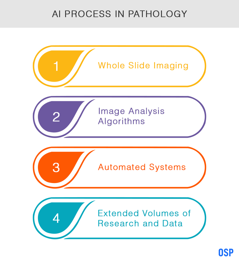
Digital Whole Slide Imaging (WSI) expands its reach by capturing the entire sample/ specimen on the slide, as opposed to the limited view of microscopic study.
The data that ensues from WSI is far more comprehensive and is recorded for further research
Pathologists in the digital realm are no longer dealing with glass, but with pixels that can be sent to any part of the world for specialized examination.
Through image analysis algorithms, AI in medical testing allows for automated analysis that is generated through the basis of research and recorded data in the software.

Broadly speaking, there are eight overarching features of digital pathology, within which artificial intelligence lends itself. They are as follows:
- Image Acquisition
- Image Storage
- Image Management
- Image Editing
- Image Manipulation
- Image Viewing
- Image Transmission
- Image Sharing
According to a published report from NCBI, “The introduction of digital pathology to nephrology provides a platform for the development of new methodologies and protocols for visual, morphometric and computer-aided assessment of renal biopsies. Application of digital imaging to pathology made substantial progress over the past decade; it is now in use for education, clinical trials and translational research. Digital pathology evolved as a valuable tool to generate comprehensive structural information in digital form, a key prerequisite for achieving precision pathology for computational biology. The application of this new technology on an international scale is driving novel methods for collaborations, providing unique opportunities but also challenges. Standardization of methods needs to be rigorously evaluated and applied at each step, from specimen processing to scanning, uploading into digital repositories, morphologic, morphometric and computer-aided assessment, data collection and analysis.”
Conclusion:
The acceptance of the advantages towards artificial intelligence in pathology are being realised, albeit slowly. Even with the compelling advantages of AI-based diagnostics and image rendition, the adoption is far from universal. There is basic scepticism towards new technologies when it comes to diagnosis and the comfort level of traditional pathologists to work with conventional models. Further, none of the above mentioned image-related processes have been nationally standardized, which is a major hurdle towards universal adaptation and routine clinical work. Through standardization of image capturing and rendition, pathologists around the globe can follow the universal steps in a consistent fashion. The NCBI publication emphasizes, “Standardization of every step in pre-analytic, analytic and post-analytic phases is crucial to achieve reliable and reproducible results across multiple groups of users.”
Another reason for hesitancy is the assumed cost involved in the implementation of the technology. However, if the advantages are weighed against the cost of artificial intelligence that latter seems to win quite easily. Laboratories also need to be upgraded, be it the storage size of their computer or the necessary equipment, for the adoption of digital pathology. However, the report further adds, “As digital imaging technology becomes more sophisticated and financially feasible, medical centers have shown increasing interest in introducing digital pathology in their daily practice. Adoption of this new technology was facilitated by advances in biomedical research, an area that has expanded to encompass biomedical engineering, computer science, image analysis and clinical informatics.”
References:
OSP is a trusted software development company that delivers bespoke solutions as per your business needs. Connect with us to hire the best talents in the industry to build enterprise-grade software.

How can we help?
Fill out the short form below or call us at (888) 846-5382
Looking for software solutions to build your product?
Let's discuss your software solutions for your product in our free development acceleration call!
Get In Touch arrow_forwardDiscuss Your Project Handover with a team of expert Book a free consultation arrow_forward
About Author

Written by Riken Shah linkedin
Riken's work motto is to help healthcare providers use technological advancements to make healthcare easily accessible to all stakeholders, from providers to patients. Under his leadership and guidance, OSP Labs has successfully developed over 600 customized software solutions for 200+ healthcare clients across continents.


